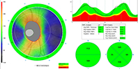
 Optovue ivue80 , Optical coherence tomography (OCT) provides high-resolution images of structures at the back of the eye, including macular, choroid, and vitreous humour. OCT utilises spectrum source of light to identify layers of structures in the eyes. We use OCT to detect pathological conditions of the retina, such as macular degeneration and glaucoma.
 Medmont topography provides both a qualitative and quantitative evaluation of corneal front curvature by using placido imaging, while the Pentacam AXL is the cutting edge of tomography, advancing beyond reflective measuring systems.
Pentacam AXL is used to measure the elevation of both the front and back surfaces of the cornea with Scheimpflug-based imaging. It also measures corneal thickness and volume, anterior chamber conformation and volume, and anterior lens topography and density also axial length. It collects data independent of tear film quality, therefore it’s much better for mapping irregular cornea, and dry eyes.
We can monitor how the cataract progresses and take preoperative measurements for IOL selection in collaboration with your Ophthalmologist. If you have had cataract surgery before, this equipment also checks that your new lens is positioned exactly where it needs to be.
We use both technologies to identify subtle corneal shape anomalies and pathological conditions, such as keratoconus and corneal ectasia. The data from the measurements can not only be used for diagnostic purposes but also used to design and simulate various custom-made contact lenses such as orthok-k lenses, corneal RGP and scleral lenses.
Corneal tomography is also used to monitor progression of keratoconus, and assess cornea condition after laser surgery or corneal transplants. The software gives the most detailed information of the cornea including the thickness and contour of the cornea , which is highly useful in monitoring conditions related to the corneal disease such as keratoconus, post laser cornea ectasia.

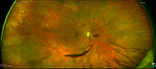
 Optos technology uses an ellipsoidal mirror in the image pathway. It is able to scan the retina all the way out to the periphery 200 degrees of retina.
Incorporates autofluorescence imaging, Optos can be used to identify a wide range of abnormalities in eyes, including age-related macular degeneration (AMD), retinal detachment, inherited dystrophies such as vitelliform lesions, central serous chorioretinopathy, choroidal tumours and nevi, inflammatory eye diseases and optic nerve head drusen. FAF is also helpful in screening for medication toxicity, including eye problems related to hydroxychloroquine (Plaquenil). FAF imaging is an easy, safe, and cheap method that is particularly useful in establishing a difficult diagnosis.
 Pentacam AXL uses high frequency sound waves (ultrasound) to measure of optical structures; including cornea, anterior chamber, lens, and vitreous chamber and axial length of the eye. It can be used to accurately calculate intraocular lens power for cataract surgery. The built-in software includes the most updated IOL formulae such as barrett II.
Since it is able to measure the length of the eye ball, it is also used to monitor myopia progression in children.
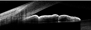
 We perform OCT on the front segment of the eye including the cornea (clear front cover of the eye), the iris (coloured circle of the eye) and the anterior angle where the cornea and iris meet at the front of the eye.
It offers a wide array of measurements that can benefit scleral lens fitting and refining scleral lens prescription.
Visual field test is used to detect glaucoma. If glaucoma is diagnosed, visual field tests are usually repeated once or twice a year to check for any changes in your vision field.
Gonioscopy exam the angle behind the pupil where the iris meets the cornea. It helps to differentiate between angle closed glaucoma and angle-open glaucoma.